
Occurrence of Stellate Ganglion: A Fetal Study
Article Sidebar
Main Article Content
Abstract
Introduction : The stellate ganglion also called as cervicothoracic ganglion, which is collection of sympathetic nerves .The ganglion lies anterior to the neck of the 1st rib.The dimensions of ganglion is about 2.5 cm in its length x1 cm in width x 0.5 cm about thickness. It might reach to anterior part of horizontal process of C7 vertebrae. The longus coli muscle is located anterolaterally ,scalene muscle lies medial to the stellate ganglion.A suprapleural membrane structurally separates the ganglion from the caudal side of the cervical pleura. The trachea, oesophagus as well as the vertebral column lies medial to stellate ganglion.
Aim : To identify stellate ganglion,its position in relation to the vertebrae .To note the size of stellate ganglion and its histology.
Materials and method: The present work was carried on 50 aborted formalin preserved human fetus specimens ranged from 11th -28th week of gestation in Department of Anatomy, GMCH , Chandigarh received in regard to autopsy. The fetuses were grouped into four age groups on the basis of gestation.Group A (11-15 weeks),Group B(15+_20 weeks),Group C(20+_25 weeks) and Group D (25 weeks onwards). The stellate ganglion was observed and its position , size was noted.
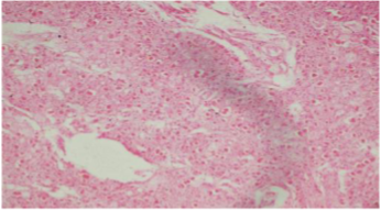
Result: Among 50 fetuses,the stellate ganglion also called as cervicothoracic ganglion were present in every age group from 11 weeks to 28 weeks of gestational age . The stellate ganglion was accounted for 64% in the fetus cervical sympathetic chains.The position of stellate ganglion at the level of C7-T1 was most common and constant.
Conclusion: The Stellate ganglia aggregation produce a complete sympathectomy to structures of head & neck region.The detailed knowledge of occurrence of stellate ganglion, its position in different gestational age groups of fetus would be guide for embryologist ,anatomist ,surgeons.
Article Details
References
GAG Decker,DJ du Plessis. Lee mcgregors. Synopsis of the autonomic nervous system .1995;212-13.
Greys H. Anatomy of human body.Philadelphia:Lea & Feibger.1918:560.
Piraccini E, Munakomi S, Chang KV. Stellate Ganglion Blocks. StatPearls Publishing; Treasure Island (FL): Aug 9, 2021.
Carron H, Litwiller R. Stellate ganglion block. Anesth Analg. 1975 Sep-Oct;54(5):567-70. [PubMed]
Wang D. Image Guidance Technologies for Interventional Pain Procedures: Ultrasound, Fluoroscopy, and CT. Curr Pain Headache Rep. 2018 Jan 26;22(1):6.
Buckley FP, MorriccaG, Murphy TM. Neurolytic blockade and hypophysectomy. In: Bonica JJ (ed). The Management of Pain, ed 2. Philadelphia, Lea and Febiger, Vol. II, 1990; 2012-14.
Raj.PP. Stellate ganglion block. In: Waldman SD, Winne AP (eds). Interventional Pain Management. Philadelphia, Dannemiller Education, WB Saunders Company, 1996.
Hogan Q, Erickson SJ. MR imaging of the stellate ganglion; Normal appearance. AJR 1992; 158:655- 659.
Grants ,JCB.An atlas of anatomy .vol II baltiomore.The Williams and wilkins co.1943;542.
Huntoon M.A.The vertebral artery is unlikely to be sole source of vascular complications occurring during stellate block.Pain Pract.2010;10:25-30.
Honma M.,Murakami G., Sato T.J., Namiki A. Spread of injectate during C6 stellate ganglion block and fascial arrangement in the prevertebral region: An experimental study using donated cadavers.Regd.Anesth.Pain Med.2000;25:573-83.
De Gama,B Z ,Lazarus L,Partab P &Satyapal, K S.The sympathetic and parasympathetic contributions to the cardiac plexus: a fetal study.Int J.Morphol.,2012; 30(4):1569-1576.
Kommuru H, Jothi S, Bapuji P, Sree D L, Antony J. Thoracic part of sympathetic chain and its branching pattern variations in South Indian cadavers. J ClinDiagn Res.2014;8(12).